
McMahon Center for Breast Health
Advanced Screening for Lifelong Breast Health
The chance of a woman having invasive breast cancer some time during her life is about 1 in 8. With odds like that, you want the best screening tool available to find, diagnose, and treat breast cancer for you or your loved one. Memorial Health System is proud to offer digital mammographies, along with our expanding state-of-the-art technologies available to diagnose and treat disease.

What is a mammogram?
A mammogram is an x-ray that gives a picture of the internal structure of the breast. There are two types of mammography: Screening and Diagnostic. Screening mammography is performed on asymptomatic women to check for undetected tumors. Diagnostic mammography is used to follow up on breast abnormalities detected during a self exam or check-up.
Digital mammograms are not widely available, but are offered at Memorial
Memorial utilizes the FCRm full-field digital mammography system, installed by Fujifilm Medical Systems. Digital mammograms are performed on a daily basis, right here in Southwest Oklahoma.
Only 8 percent of mammography units in the United States are digital. Digital mammography is a relatively new technology and there are currently a small number of facilities with digital units. A recent study of about 50,000 women found that digital mammograms were actually better than film mammograms in picking up cancers in certain groups of women, including women with particularly dense breasts and women younger than 50. Researchers aren’t sure why digital is more accurate for these women, but they think it’s because digital photos can be manipulated to get a better view.
Why mammogram?
Mammography is the leading defense against breast cancer. Memorial offers digital mammograms, a procedure capturing images of the breast image special electronic x-ray detector, which converts the image into a digital picture for review on a computer monitor. With digital mammography, the magnification, orientation, brightness, and contrast of the image may be altered after the exam is completed to help the radiologist more clearly see certain areas.
Benefits of Digital Mammograms
- Improved contrast between the dense and non-dense breast tissue
- Faster image acquisition (less than a minute)
- Shorter exam time (approximately half that of film-based mammography)
- Easier image storage
- Physician manipulation of breast images for more accurate detection of breast cancer
- Ability to correct under or over-exposure of films without having to repeat mammograms
What to expect during your mammography
First, you will be asked to undress from the waist up. A female Radiologic Technologist certified by the American Registry of Radiological Technologists and specializing in mammography will perform the procedure. The technologist will then place your breast between two plates and compress them to take the x-ray. With your comfort in mind, we use Mammo Pads, single-use foam cushions, which offer a warmer and softer barrier between the surfaces and edges of the mammography device. The experience is more comfortable, without impacting the clarity of the mammogram. Once the procedure is completed the exam is then sent through the iCAD computer-aided detection to provide a secondary computer-aided interpretation of any areas of concern within the breast. A board-certified radiologist will interpret your exam and a report will be sent to your physician, and a patient result letter will be sent to you.
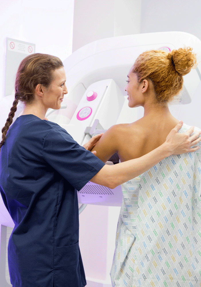
How Does Digital Mammography Differ From Standard Mammography?
In standard mammography, images are recorded on film using an x-ray cassette. The film is viewed by the radiologist using a “lightbox” and then stored in a jacket in the facility’s archives. With digital mammography, the breast image is captured using a special electronic x-ray detector, which converts the image into a digital picture for review on a computer monitor. The digital mammogram is then stored on a computer. With digital mammography, the magnification, orientation, brightness, and contrast of the image may be altered after the exam is completed to help the radiologist more clearly see certain areas.
Studies have shown that digital mammography may provide additional benefits, such as lower radiation doses and higher sensitivity to abnormalities. Other data from German researchers suggest that the radiation dose can be reduced by up to 50% with digital mammography and still detect breast cancer as well as the standard radiation dose of film mammography.
It is suspected that digital mammography is superior in some women because it allows radiologists to heighten the contrast between dense tissue and cancerous cells, which both appear to be white on a film mammogram.
Conventional film mammography requires several minutes to develop the film while digital mammography provides the image on the computer monitor in less than a minute after the exposure/data acquisition. Thus, digital mammography provides a shorter exam for the woman. Digital mammography can also be manipulated to correct for under or overexposure after the exam is completed, eliminating the need for some women to undergo repeat mammograms before leaving the facility.
The Women’s Imaging facility at Memorial is certified by the American College of Radiology and the Mammography Quality Standards Act. It is also licensed by the Oklahoma Department of Public Health and is a Softer Mammogram provider.
Breast Imaging Beyond Mammography
Taking charge of her health is one of the smartest things a woman can do for herself. When the need for imaging services arises, Memorial is the best choice in Southwest Oklahoma.
Women’s Imaging Services offers women the latest in technological advances including digital mammography services, the ABBI Breast Biopsy System, and bone densitometry.
Pre-Operative Hook-Wire Localization
Hook-wire localization is a mammographically-guided procedure performed to help guide a surgeon to a lump that will be surgically excised. It is done immediately before a surgical biopsy. This procedure is minimally painful and is associated with very minimal risks.
Breast Ultrasound
A follow up to further assess a lump found on mammography using sound waves to produce an image of the internal structures.
Stereotactic Breast Biopsy
A procedure that offers a minimally invasive way of diagnosing a suspicious finding. To undergo a stereotactic breast biopsy, a woman is given a local anesthetic on the impacted skin and placed on her stomach in a special mammography unit. A computer assists your surgeon in guiding a special biopsy needle into the suspicious area using digital imaging guidance. Very small core biopsies of the breast are taken. The procedure is brief, minimally painful. The removed tissue is sent to pathology for a complete evaluation.
Risk Factors for Breast Cancer include:
-
A family history of breast cancer (particularly a mother, sister or daughter)
-
A previous diagnosis of cancer in one breast
-
A prior abnormal breast biopsy
-
Menstruation before 12 and menopause after 55
-
Giving birth later in life or being childless
-
Taking hormone replacement therapy
-
Having dense breast tissue
How do I make an Appointment?
To schedule a screening mammogram, please call 580.250.5846.
If you need a diagnostic mammogram please have your physician set up your appointment. For added convenience, your physician can schedule your bone density screening and mammogram back-to-back on the same day. An order from your physician will be needed for the diagnostic mammogram and bone density test.
Contact Us

Phone

Address
3201 W Gore Blvd
Lawton, OK 73505