Cardiovascular and Thoracic Surgery
Surgical Procedures
Learn more about each of the following procedures by clicking on the plus sign.

Heart
Our surgeons utilize the least invasive techniques possible to achieve less blood loss, reduce pain, and achieve a shorter length of stay and faster recovery.
Coronary Artery Bypass Graft (CABG)
Coronary artery bypass graft (CABG) is surgery to treat coronary artery disease. The surgery helps blood make a detour, or bypass, around one or more narrowed or blocked coronary arteries. Coronary arteries are the blood vessels that bring blood to the heart. The surgery is also called coronary artery bypass or bypass surgery. Your physician will make a bypass using a piece of blood vessel from another part of your body. Your physician will attach, or graft, this blood vessel above and below the narrowed or blocked section of your artery. The most common way to do bypass surgery is through a large cut, called an incision, in the chest. This is called open-chest surgery. Your physician will make the cut in the skin over your breastbone (sternum). Then the physician will cut through your sternum to reach your heart and coronary arteries. The physician will connect you to a heart-lung bypass machine. This machine will let the physician stop your heart while he or she works. The physician will use a blood vessel from your chest, arm, or leg to bypass the narrowed or blocked arteries. When the blood vessels are in place, the physician will restart your heart. The physician will use wire to put your sternum back together. The wire will stay in your chest. You will get stitches or staples to close the cuts in your skin. The cuts will leave scars that may fade in time.
View our full informational packet.
Off-Pump Coronary Artery Bypass (OPCAB)
Off-Pump Coronary artery bypass graft is surgery to treat coronary artery disease. This procedure is performed without using the heart-lung machine. The surgery helps blood make a detour, or bypass, around one or more narrowed or blocked coronary arteries. Coronary arteries are the blood vessels that bring blood to the heart. The surgery is also called coronary artery bypass or bypass surgery. Your physician will make a bypass using a piece of blood vessel from another part of your body. Your physician will attach, or graft, this blood vessel above and below the narrowed or blocked section of your artery while the heart is still beating.
Mitral Valve Replacement
Mitral valve surgery can repair or replace your heart’s mitral valve. The new valve may be mechanical or made of animal tissue, often from a pig. Your physician will talk with you about which type of valve is best for you.
The mitral valve opens and closes to keep blood flowing in the proper direction through your heart. When the mitral valve does not close properly, it’s called mitral valve regurgitation. If the valve is very tight and narrow, it’s called mitral valve stenosis. In both of these cases, blood does not flow through the heart the right way.
The physician will make a cut in the skin over your breastbone (sternum). This cut is called an incision. Then the physician will cut through your sternum to reach your heart. The physician will connect you to a heart-lung bypass machine. It adds oxygen to your blood and moves the blood through your body. This machine will allow the physician to stop your heartbeat while he or she works on your heart. After the physician has repaired or replaced your mitral valve, he or she will restart your heartbeat. Then the physician will use wire to put your sternum back together. Your incision will be closed with stitches or staples. The wire will stay in your chest. The incision will leave a scar that will fade with time. You will stay in the hospital for 3 to 8 days after surgery.
View our full informational packet.
Aortic Valve Replacement
Aortic valve replacement gives you a new aortic heart valve. The new valve may be mechanical or made of animal tissue, often from a pig. Your physician will talk with you before surgery about which type of valve is best for you. The aortic valve opens and closes to keep blood flowing in the proper direction through your heart. When the aortic valve does not close properly, it’s called aortic valve regurgitation. If the valve is very tight and narrow, it’s called aortic valve stenosis. In both of these cases, blood does not flow through the heart the right way. You will be asleep during the surgery. Your physician will make a cut in the skin over your breastbone (sternum). This cut is called an incision. Then the physician will cut through your sternum to reach your heart.
The physician will connect you to a heart-lung bypass machine. It adds oxygen to your blood and moves the blood through your body. This machine will allow the physician to stop your heartbeat and replace the valve.
After the physician has replaced your aortic valve, he or she will restart your heartbeat. Then the physician will use wire to put your sternum back together. Your incision will be closed with stitches or staples. The wire will stay in your chest. The incision will leave a scar that may fade with time.
You will stay in the hospital for 3 to 8 days after surgery.
View our full informational packet.
Surgical Treatment of Atrial Fibrillation
Atrial fibrillation (say “AY-tree-uhl fih-bruh-LAY-shun”) is the most common type of irregular heartbeat (arrhythmia). Normally, the heart beats in a strong, steady rhythm. In atrial fibrillation, a problem with the heart’s electrical system causes the two upper parts of the heart (the atria) to quiver, or fibrillate. Your heart rate also may be faster than normal. Atrial fibrillation can be dangerous because if the heartbeat isn’t strong and steady, blood can collect, or pool, in the atria. And pooled blood is more likely to form clots. Clots can travel to the brain, block blood flow, and cause a stroke. Atrial fibrillation can also lead to heart failure. Treatment for atrial fibrillation helps prevent stroke and heart failure. It also helps relieve symptoms. Atrial fibrillation is often caused by another heart problem. It may happen after heart surgery. It may also be caused by other problems, such as an overactive thyroid gland or lung disease. Many people with atrial fibrillation are able to live full and active lives.
Chest
Our surgeons partner with colleagues and referring physicians to ensure rapid access and timely evaluation for new patients and consultations.
Conditions Treated
- Early and locally advanced lung cancer
- Pretreatment staging of thoracic neoplasms
- Lung Volume Reduction Surgery
- Tracheal tumors and airway management
- Malignant pleural effusions
- Mesothelioma and other malignant pleural diseases
- Chest wall tumors and chest wall deformities
- Pulmonary metastases
- Mediastinal tumors
Thyroidectomy
A thyroidectomy is surgery to take out your thyroid gland. This gland is shaped like a butterfly. It lies across the windpipe (trachea). The gland makes hormones that control how your body makes and uses energy (metabolism). A physician removes the gland when it gets too big, does not work right, or has a tumor. Most tumors that grow in this gland are benign. This means they are not cancer. The physician will take out the thyroid through a cut (incision) in the front of your neck. You will likely have a tube, called a drain, in your neck to let fluid out of the cut. The drain is most often taken out before you go home. You may go home on the same day. Or you may stay one or more nights in the hospital. You may return to work or your normal routine in 1 to 2 weeks. This depends on whether you need more treatment and how you feel. It may also depend on the kind of work you do. Your physician will check your incision in about a week. You may need to take thyroid medicine. If you have thyroid cancer, you may need to have radioactive iodine therapy. Your physician will talk to you about what happens next.
View our full informational packet.
Video Assisted Thoracic Surgery
Video Assisted Thoracic Surgery or VATS is a way to do surgery inside the chest. With open surgery, the physician makes one large cut in your chest. But with VATS, the physician makes several small cuts. VATS also differs from open surgery because the physician does not have to cut through the ribs or breastbone (sternum). The physician can use VATS to find and treat many problems in the chest. To start, the physician will make several small cuts between your ribs. These cuts are called incisions. The physician will put a thin, lighted tube with a camera on it into your chest. This tube is called a thoracoscope, or scope. It lets the physician see inside your chest. Then the physician will use tiny surgical tools to do the surgery. The physician will close the incisions with stitches or staples. How long you stay in the hospital and how long your recovery takes will depend on why you are having the surgery. The scars from the incisions will fade with time. The area around the incisions may ache or feel numb in the weeks after surgery.
View our full informational packet.
Thoracotomy
A thoracotomy (say “thor-uh-KAW-tuh-mee”) is surgery done through a cut in the chest wall. The cut is called an incision. It is made between the ribs. The physician can operate inside the chest through this incision. A thoracotomy may be used for surgery on the lungs, esophagus, trachea, heart, aorta, or diaphragm. The exact place where the incision is made depends on the reason for the surgery. It is usually across the side of the mid-chest. Your physician may need to cut through a rib or the breastbone (sternum). This is done to spread the ribs far enough to do the surgery. When the surgery is finished, the physician will close the incision with stitches or staples. If a rib or the breastbone was cut, the physician will use wire to hold the pieces of bone together as they heal. Most people spend 3 to 7 days in the hospital after this type of surgery. You will be quite sore. You’ll get medicine to help with this. Even though you will be sore, make sure that you breathe deeply and are as active as you can after surgery. This will help your lungs expand again. It will help you heal more quickly too. Also, be sure not to smoke after surgery. The amount of time you will need to recover at home depends on the type of surgery you had. You will probably need to take at least 1 to 2 months off work.
Bronchoscopy
Bronchoscopy (say “bron-KOSS-koh-pee”) is a type of procedure. Your physician uses a flexible tube to look at your airway. This tube is called a bronchoscope. It lets your physician see your throat, voice box (larynx), windpipe (trachea), and bronchial tubes. There are many reasons to have this procedure. Your physician may look for problems with your airway. Or he or she may remove an object or growth. Your physician could also take a sample of tissue to study. This is called a biopsy. You will probably be awake for the procedure. But you will get medicine so you will not have pain. The physician puts the bronchoscope into your mouth or nose and down your throat. Most people go home the same day. You will probably be able to go back to work or your normal routine in 1 or 2 days.
View our fulll informational packet.
Thoracentesis
Thoracentesis (say “thor-uh-sen-TEE-sis”) is a procedure to remove fluid from the space between the lungs and the chest wall. This is called the pleural space. The procedure may also be called a “chest tap.” It is normal to have a small amount of fluid in the pleural space. But too much fluid can build up because of problems such as infection, heart failure, and lung cancer. The procedure may be done to help with shortness of breath and pain caused by the fluid buildup. Or you may have it done so the physician can test the fluid to find the cause of the buildup. Your physician will put a long, thin needle or a thin plastic tube, called a catheter, between two of your ribs. The physician will use the needle or catheter to take fluid out. You may get medicine before the procedure. This helps with pain and helps you relax. The procedure will take about 15 minutes. Most people go home shortly after. You can go back to work or your normal activities as soon as you feel up to it. If the physician sends the fluid to a lab for testing, it usually takes a few hours to get the results. Some of the test results may take a few days. The physician or nurse will discuss the results with you.
View our fulll informational packet.
Esophageal Carcinoma
An esophagectomy (say “ee-sof-uh-JEK-tuh-mee”) is surgery to remove all or part of the esophagus. The esophagus is the tube that carries food from the throat to the stomach. Nearby lymph nodes and other tissue may also be removed. The physician connects the remaining healthy part of the esophagus to the stomach or colon so that you can still swallow and eat food. This surgery is mainly used to treat esophageal cancer. But it can also be used for other problems with the esophagus. During the surgery, the physician makes large cuts in the belly and the upper chest or neck. These cuts are called incisions. Where they are depends on where the problem is. After surgery, you may be in the hospital for 1 to 2 weeks. You have to be very careful about what you eat for 1 to 2 months after surgery and maybe for the rest of your life. You may have a feeding tube (J-tube) in your belly when you go home. This is to be sure you get enough nutrition. This will come out when you are able to eat normally. How long this takes varies.
For many people, it takes 4 to 6 weeks after surgery. Your physician will give you detailed information on your diet and how to use the J-tube. You may feel weak after surgery. Take good care of yourself. Get enough rest and stay active. This can help you heal faster. You will probably need to take at least 6 to 12 weeks off from work. This depends on the type of work you do and how you feel. You will need more time to get better if you need other treatment for cancer, such as chemotherapy.
Aortic
At the base of the heart, starting at the aortic valve, branches to heart, brain, arms, spinal cord, visceral organs, kidneys, and legs originate from the aorta.
Aortic Procedures
- Aortic root aneurysms
- Ascending aortic aneurysms
- Transverse aortic arch aneurysms
- Descending thoracic aortic aneurysms
- Abdominal aortic aneurysms
- Acute and chronic aortic dissections
- Atheromatous disease of the aorta
- Coarctation
- Bicuspid aortic valve
Thoracic Endovascular Aortic Repair (TEVAR)
Aortic aneurysm repair is surgery to fix a weak and bulging section of the aorta. The aorta is the large blood vessel (artery) that carries blood from the heart through the chest and belly to the rest of the body. An aortic aneurysm that is not repaired may burst (rupture). This can be life-threatening. Your physician will use a man-made tube to replace the weak section of your aorta in your chest. The tube is called a graft. The physician will make a large cut in your chest. This cut is called an incision. It may be made through the breastbone (sternum). Or it may be in the side of your chest between your ribs. During the surgery, the physician may connect you to a machine that does the jobs of your heart and lungs. It’s called a heart-lung bypass machine. This machine lets the physician stop your heartbeat while he or she works on your aorta. The physician will put clamps on the aorta above and below the aneurysm. This stops blood flow through the area that the physician is working on. The physician will replace the weak section of your aorta with a graft. In some cases, the physician can do surgery without using a heart-lung bypass machine. After the aorta is fixed, the physician will remove the clamps. Blood can then flow through the aorta again. Then the physician will use stitches or surgical staples to close the incision in your chest. You will probably spend 5 to 7 days in the hospital. You will need to take it easy for at least 4 to 6 weeks at home.
View our full informational packet.
Open Abdominal Aortic Aneurysm Repair (AAA)
Abdominal aortic aneurysm repair is a type of surgery. It fixes an aneurysm in your aorta. An aneurysm is a weak or bulging part of a vein or artery. Your aorta is a large artery that carries blood from your heart through your belly to the rest of your body. Without surgery to fix this problem, your aorta could burst. This can cause death. To do the surgery, the physician makes a large cut in your belly. This cut is called an incision. Then the physician puts clamps above and below the weak part of the aorta. This stops blood flow. It allows the physician to replace the weak part with a tube called a graft. After the graft is in place, the physician removes the clamps so blood can flow again. Then the physician uses stitches or staples to close the incision. You will probably spend 5 to 7 days in the hospital. You will need to take it easy for at least 4 to 6 weeks at home.
View our full informational packet.
Endovascular Abdominal Aneurysm Repair (EVAR)
Endovascular aortic aneurysm repair fixes an aneurysm in your aorta. An aneurysm is a weak or bulging part of a vein or artery. Your aorta is a large artery. It carries blood from your heart through your belly to the rest of your body. If you don’t fix this problem, your aorta could burst. And this can cause death. Your physician will use a special man-made tube to fix your aorta. This is called a stent graft. After the procedure, your blood will flow through the stent graft. It will not push on the aneurysm. To do the procedure, the physician makes two cuts in your groin area. These are called incisions. Then the physician puts small tubes into the arteries in that area. The tubes are called catheters. The physician first uses the catheters to put dye in your arteries. The dye makes your aorta show up on X-rays. Next, the physician uses wires inside the catheters to move the stent graft through the arteries and up to your aorta. After the stent graft is in place, the physician takes out the catheters and wires. Then he or she uses stitches to close the incisions. You will have scars that fade with time. You will probably spend 1 to 3 days in the hospital. You may be able to return to work and many of your daily activities 1 to 2 weeks after the procedure.
View our full informational packet.
Vascular & Endovascular
Our surgeons offer a full spectrum of open and minimally invasive procedures that are tailored to patients’ needs.
Absorbable Stent
The Absorb dissolving heart stent is the first and only device of its kind – a drug-eluting coronary stent that dissolves, completely and naturally, in the body over time. Absorb treats coronary artery disease like a standard metallic stent, propping the diseased vessel open to restore blood flow, but then disappears after the artery is healed, leaving no metal behind to restrict natural vessel motion.
View our full informational packet.
Carotid Endarterectomy
A carotid endarterectomy (say “kuh-RAW-tid en-dar-tuh-REK-tuh-mee”) is done to remove fatty buildup (plaque) from one of the carotid arteries. There are two of these arteries. One runs along each side of the neck. They supply blood to your brain. When plaque builds up in either one, it can make it hard for blood to flow to the brain. This surgery may lower your risk of stroke. The physician will make a cut (incision) in your neck. Then the physician will make a cut in the carotid artery and take out the plaque. Next, the physician will close the cut in the artery with stitches. Or the physician may sew a man-made patch over this cut. This will make the artery wider. It also helps keep it from getting narrow again. Then the physician will use stitches to close the cut in your skin. It will leave a scar. But the scar will fade with time. You will probably go home the day after surgery. You may be able to go back to work or your usual activities in 1 to 2 weeks.
View our full informational packet.
Femoral to Popliteal Bypass
A femoropopliteal bypass is a type of surgery. It is also called a fem-pop or leg artery bypass. This type of surgery redirects blood around a blocked blood vessel in your leg. So it may improve the blood flow in your leg. This can decrease leg pain, numbness, and cramping. Your physician will use something called a graft to make the blood go around (bypass) the blocked part of your blood vessel. The graft may be a vein taken from another place in your leg. Or it may be a man-made blood vessel. You will probably be asleep during the surgery. But it also can be done while you are awake. If you are awake, you will get medicine to numb your leg and prevent pain. First, the physician makes cuts in your thigh and sometimes in the side of the calf. These cuts are called incisions. If one of your veins is being used for the graft, the physician will make other incisions in your leg. Then the physician will attach one end of the graft to the femoral artery in your thigh. The other end will be attached to the popliteal artery above or below your knee. After the graft is in place and blood is flowing through it, the physician uses stitches or staples to close the incisions. You will have scars, but they will fade with time. You will probably spend 2 to 4 days in the hospital. For at least 2 to 6 weeks, you will need to take it easy at home. It may take 6 to 12 weeks to fully recover.
View our full informational packet.
Below Knee Leg Amputation
A below-the-knee amputation is surgery to remove your leg below the knee. Your physician removes the leg and keeps as much healthy skin, blood vessel, and nerve tissue as possible. Having your leg removed is traumatic. You have to learn to live with new limitations. This can be hard and frustrating. You may feel depressed. Or you may grieve for your previous lifestyle. Talking with your family, friends, and health professionals about how you feel may help. You may also find it helps to talk with a person who has had an amputation. Even though losing a limb is a challenge, it does not change who you are. It doesn’t prevent you from enjoying life. You will have to learn new ways to do things. But you will still be able to work and take part in sports and activities. And you can still learn, love, play, and live life to its fullest. Your physician will tell you how much of your leg should be removed. He or she will leave enough healthy skin to cover the remaining part of your leg (residual limb). You may get an artificial leg. This is called a prosthesis. If you get one, your physician will shape your residual limb for the best possible fit. Your physician may sew together the skin to cover the residual limb. Or he or she may leave it open to make sure it heals as it should. In this case, the skin may be sewn together 10 to 14 days later. After surgery, you will stay in the hospital for several days. How long you stay depends on your general health and the way your physician does the surgery. You may spend part of your recovery in a skilled nursing facility. Your residual limb may heal as soon as 4 to 8 weeks after surgery. But it may take longer. You will need physical rehabilitation (rehab). The rehab can sometimes start within 48 hours of your surgery. It may last as long as 1 year.
View our full informational packet.
Foot Amputation
Foot amputation is surgery to remove part or all of your foot. Your physician will leave as much healthy skin, blood vessel, and nerve tissue as possible. You will be asleep during the surgery. Your physician will tell you how much of your foot should be removed. He or she will leave enough healthy skin to cover the residual limb or the remaining part of your foot. Some people get an artificial foot. This is called a prosthesis. If you get one, your physician will shape the remaining part of your leg or foot for the best possible fit. Your physician may sew the skin closed to cover the residual limb or remaining part of your foot. Or he or she may leave it open to make sure that it heals as it should. In this case, the skin may be sewn together several days later. Or it may be left open to heal on its own. Skin that is left open can take a few months to close. How long you will stay in the hospital after surgery depends on how much of your foot was removed. It also depends on your general health. You may need physical rehabilitation (rehab) after the surgery. Rehab can sometimes start within 48 hours of your surgery. It may last as long as 1 year. Having part or all of your foot removed is traumatic. Learning to live with new limitations can be hard and frustrating. You may feel depressed. Or you may grieve for the lifestyle you used to have. Talking with your family, friends, and health professionals about your frustrations may help. You may also find that it helps to talk with a person who has had an amputation. Remember that even though losing part or all of your foot is a challenge, it does not change who you are or prevent you from enjoying life. You will have to adapt and learn new ways to do things. But you will still be able to work and take part in sports and activities. And you can still learn, love, play, and live life to its fullest.
View our full informational packet.
Varicose Veins
Varicose veins are twisted, enlarged veins near the surface of the skin. They develop most often in the legs and ankles. Some people may be more likely than others to get varicose veins because of aging or hormone changes or because a parent has them. Being overweight or pregnant can make varicose veins worse. Jobs that require standing for long periods of time also can make them worse.
View our full informational packet.
Microphlebectomy
Microphlebectomy (say “my-kroh-fluh-BEK-tuh-mee”) is a procedure used to remove varicose veins. These are twisted and enlarged veins near the surface of the skin. The procedure is also called ambulatory phlebectomy or stab avulsion. The procedure is usually done in your physician’s office. You will get medicine to make you relax or to numb the area. Your physician will make several tiny cuts (incisions) in the skin. The varicose veins will be removed through the cuts. You most likely will not need stitches to close the cuts. It usually takes less than 1 hour.
Femoral-Tibial Bypass
Femoral-tibial bypass is a type of surgery. It redirects blood around blocked blood vessels in your lower leg or foot. It is often done if you have pain. Or it may be done if you have foot sores caused by circulation problems. Your physician will use something called a graft to make the blood go around (bypass) the blocked part of your blood vessel. Often the graft is a vein taken from another place in your leg. But sometimes it is a man-made blood vessel. The graft will carry blood from the femoral artery in your groin to the tibial artery in your lower leg or foot. You will be asleep during the surgery. Or you will get medicine to numb your lower body and prevent pain. The physician will make cuts in your skin above and below the blocked blood vessel. These cuts are called incisions. If one of your veins is being used for the graft, the physician will make another incision in your leg to take out the vein. Then the physician will attach one end of the graft to the femoral artery and the other end to the tibial artery. After the graft is in place and blood is flowing through it, the physician will use stitches or staples to close the incisions. You will have scars, but they will fade with time. You may need to stay in the hospital for 3 to 5 days. It may take 6 to 12 weeks to fully recover.
View our full informational packet.
Aortobifemoral Bypass
An aortobifemoral bypass is surgery to move blood flow around blocked blood vessels in your belly or groin area. This will increase blood flow to your legs. It may allow you to walk farther. The physician will use a blood vessel, called a graft, to bypass the blocked blood vessels. The graft will carry blood from the aorta to the femoral artery. This artery is in the groin area of each thigh. The aorta is the large blood vessel that carries blood from the heart to the blood vessels in the belly. The femoral arteries are large blood vessels that carry blood from the blood vessels in the belly to the legs. The physician will make a cut in your belly. He or she will also make a cut in your groin at the top of each thigh. These cuts are called incisions. The physician will put the graft in your belly through the incisions. The graft is connected to the aorta and the femoral arteries. Stitches or staples are used to close the incisions. You will probably spend 4 to 7 days in the hospital. You will need to take it easy for at least 4 to 6 weeks at home.
View our full informational packet.
Peripheral Arterial Disease (PAD)
Peripheral arterial disease (PAD) is narrowing or blockage of arteries in your arms and legs. The most common cause of PAD is the buildup of plaque on the inside of arteries. Plaque is made of extra cholesterol, calcium, and other material in your blood. Over time, plaque builds up in the walls of the arteries, including those that supply blood to your legs. This buildup leads to poor blood flow. When you have PAD, you have a risk of having plaque in other arteries in your body. This raises your risk of a heart attack and stroke. This information focuses on peripheral arterial disease of the legs, the area where it is most common. When you have PAD of the legs and you walk or exercise, your leg muscles do not get enough blood, and you can get painful cramps. The cramps are called intermittent claudication. Peripheral arterial disease is also called peripheral vascular disease.
View our full informational packet.
Above Knee Leg Amputation
An above-the-knee amputation is surgery to remove your leg above the knee. Your physician removes the leg and keeps as much healthy skin, blood vessel, and nerve tissue as possible. Having your leg removed is traumatic. You have to learn to live with new limitations. This can be hard and frustrating. You may feel depressed. Or you may grieve for your previous lifestyle. Talking with your family, friends, and health professionals about how you feel may help. You may also find it helps to talk with a person who has had an amputation. Even though losing a limb is a challenge, it does not change who you are. It doesn’t prevent you from enjoying life. You will have to learn new ways to do things. But you will still be able to work and take part in sports and activities. And you can still learn, love, play, and live life to its fullest. Your physician will tell you how much of your leg should be removed. He or she will leave enough healthy skin to cover the remaining part of your leg (residual limb). You may get an artificial leg. This is called a prosthesis. If you get one, your physician will shape your residual limb for the best possible fit. Your physician may sew together the skin to cover the residual limb. Or he or she may leave it open to make sure it heals as it should. In this case, the skin may be sewn together 10 to 14 days later. After surgery, you will stay in the hospital for several days. How long you stay depends on your general health and the way your physician does the surgery. You may spend part of your recovery in a skilled nursing facility. Your residual limb may heal as soon as 4 to 8 weeks after surgery. But it may take longer. You will need physical rehabilitation (rehab). The rehab can sometimes start within 48 hours of your surgery. It may last as long as 1 year.
View our full informational packet.
Toe Amputation
A toe amputation is surgery to remove one or more toes. You will get medicine to help you relax and numb your foot. Then your physician will make a cut (incision) to remove your toe. If you have healthy skin to cover the wound and have no signs of infection, the physician will then try to close the wound. You may have problems with walking or balance, especially if you lose your big toe. But you will not need an artificial toe. You may need to have special insoles made to fit in your shoes. These insoles are called orthotics. You may go home on the day of surgery. Or you may need to stay in the hospital for a few days. This depends on how fast you recover and on your overall health.
View our full informational packet.
Sclerotherapy
Sclerotherapy is a treatment to get rid of varicose veins. A chemical called a sclerosant is injected into the varicose vein. This causes the vein to close. The procedure may cause some pain. The chemical may cause burning or cramping for a few minutes at the injection site. The procedure can take up to 30 minutes. It depends on how many veins are treated and how big they are. You should be able to walk on your own after the treatment.
View our full informational packet.
Endovenous Ablation
Endovenous ablation is a procedure to close off varicose veins. Endovenous means that the procedure is done inside the vein. Ablation means a physician uses heat to damage and close off the vein. Varicose veins are twisted, enlarged veins near the surface of the skin. The heat damages a vein, and scar tissue forms. This scar tissue closes the vein. A closed vein loses its source of blood and dies. Eventually, you won’t be able to see the veins anymore. The heat used for ablation can come from a laser or radio waves. A laser is a highly focused beam of light. Certain radio waves can make energy and heat called radiofrequency energy.
View our full informational packet.
Meet Our Providers

Colleen Gallimore
PA-C
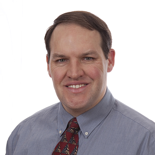
Aaron L. Trachte
MD
Contact Us

Memorial Health Heart & Vascular
Memorial Health Cardiovascular & Thoracic Surgery